Home » Without Label » Anterior Muscles Of The Body Labeled - Muscle chart with most important muscles of the human body ... - Labeled anterior and posterior muscles of the body.
Anterior Muscles Of The Body Labeled - Muscle chart with most important muscles of the human body ... - Labeled anterior and posterior muscles of the body.
Anterior Muscles Of The Body Labeled - Muscle chart with most important muscles of the human body ... - Labeled anterior and posterior muscles of the body.. Arm anterior 3d illustration project. Anatomynote.com found main muscles of human body anterior view from plenty of anatomical pictures on the internet. Muscle tone provides a slight tension on the muscle to prevent damage to the muscle and joints from sudden movements, and also helps to maintain the body's posture. Jul 26, 2021 · iliopsoas acts as the antagonist of the gluteus maximus muscle and the hamstring. Muscle anatomy quiz for anatomy and physiology!
This quiz requires labeling, so it will test your knowledge on how to identify these muscles (latissimus dorsi, trapezius, deltoid, biceps brachii, triceps brachii, brachioradialis, pectoralis major, serratus anterior, rectus abdominis, etc.). This is a table of skeletal muscles of the human anatomy. This image added by admin. Deep to the gluteus maximus is the gluteus medius, and deep. Thank you for visit anatomynote.com.
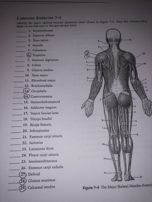
Solved: LABELING EXERCISE 7-4 Ldenify The Major Skeletal M ... from media.cheggcdn.com We think this is the most useful anatomy picture that you need. Choose from 500 different sets of muscles of the body anterior flashcards on quizlet. The anterior serratus pulls the scapula outward which lifts the shoulder. Muscle anatomy quiz for anatomy and physiology! Label, name the muscle group. Serratus anterior superior, serratus anterior intermediate, serratus anterior inferior and runs from the front of the chest around the side to the scapula. The anterior and middle scalene muscles, which also are located at the sides of the neck, act ipsilaterally to rotate the neck, as well as to elevate the first rib. Serratus anterior this muscle is divided into three named parts:
Thank you for visit anatomynote.com.
Almost every muscle constitutes one part of a pair of identical bilateral muscles, found on both sides, resulting in approximately 320 pairs of muscles, as presented in this article. Learn vocabulary, terms, and more with flashcards, games, and other study tools. This is an online quiz called muscles of the anterior surface of the body. In the anterior compartment, they are split into three categories: The anterior and middle scalene muscles, which also are located at the sides of the neck, act ipsilaterally to rotate the neck, as well as to elevate the first rib. Labeled anterior and posterior muscles of the body. Its primary job is to rotate and flex the bones of the neck unilaterally (using only one muscle of the pair), as well as to. See more ideas about muscle anatomy, human anatomy and physiology, body anatomy. We think this is the most useful anatomy picture that you need. Muscle anatomy quiz for anatomy and physiology! You can click the image to magnify if you cannot see clearly. This quiz requires labeling, so it will test your knowledge on how to identify these muscles (latissimus dorsi, trapezius, deltoid, biceps brachii, triceps brachii, brachioradialis, pectoralis major, serratus anterior, rectus abdominis, etc.). The reason for this is their origin at specific points on the tibia or fibula and insertion on certain areas.
Side bending also is an important action of the cervical spine. You've just got five to. The posterior tibial allows the foot to extend. All muscles maintain some amount of muscle tone at all times, unless the muscle has been disconnected from the central nervous system due to nerve damage. This article will cover the attachments, actions, innervations and clinical correlations of these muscles.
labeled posterior thigh muscles | Anatomy images, Body ... from i.pinimg.com Learn muscles of the body anterior with free interactive flashcards. Flexor carpi ulnaris is a fusiform muscle located in the anterior compartment of the forearm. All muscles maintain some amount of muscle tone at all times, unless the muscle has been disconnected from the central nervous system due to nerve damage. See more ideas about muscle anatomy, human anatomy and physiology, body anatomy. Browse or search in thousands of pages or create your own page using a simple wizard. Together, these muscles straighten your knee, stabilize your knee joint, assist in flexing your hip (drawing your knee towards your chest), and help absorb force when you land after jumping or leaping. This is a table of skeletal muscles of the human anatomy. Learn vocabulary, terms, and more with flashcards, games, and other study tools.
Make writing personal training programs easy with these custom designed exercise templates, and keep your clients focused and progressing.
This image added by admin. The scalenus anterior muscle is a muscle of inspiration (breathing in). There are around 650 skeletal muscles within the typical human body. It also supports the plantar arch. Flexor carpi ulnaris is a fusiform muscle located in the anterior compartment of the forearm. These are the rectus abdominis, pyramidalis, external abdominal oblique, internal abdominal oblique and transversus abdominis. Muscular system anterior labeled.the muscles labelled in the anterior muscles diagram shown above are listed in bold in the following table sternocleidomastoid trapezius serratus anterior upper body muscles labeled major anterior muscles. The anterior and middle scalene muscles, which also are located at the sides of the neck, act ipsilaterally to rotate the neck, as well as to elevate the first rib. The psoas major and iliacus make up the iliopsoas group.some of the largest and most powerful muscles in the body are the gluteal muscles or gluteal group.the gluteus maximus is the largest; You've just got five to. This quiz requires labeling, so it will test your knowledge on how to identify these muscles (latissimus dorsi, trapezius, deltoid, biceps brachii, triceps brachii, brachioradialis, pectoralis major, serratus anterior, rectus abdominis, etc.). Together, these muscles straighten your knee, stabilize your knee joint, assist in flexing your hip (drawing your knee towards your chest), and help absorb force when you land after jumping or leaping. Labeled anterior and posterior muscles of the body.
The superficial muscles in the anterior compartment are the flexor carpi ulnaris, palmaris longus, flexor carpi radialis. This article will cover the attachments, actions, innervations and clinical correlations of these muscles. Gluteal region muscles that move the femur. Click on the tags below to find other quizzes on the same subject. Muscle tone provides a slight tension on the muscle to prevent damage to the muscle and joints from sudden movements, and also helps to maintain the body's posture.
Muscle and Tendon Characteristics - Classic Human Anatomy ... from schoolbag.info This is a table of skeletal muscles of the human anatomy. Choose from 500 different sets of muscles of the body anterior flashcards on quizlet. Gluteal region muscles that move the femur. Nevertheless, the exact number is difficult to define. Flexor carpi ulnaris is a fusiform muscle located in the anterior compartment of the forearm. The posterior tibial allows the foot to extend. This is an online quiz called muscles of the anterior surface of the body. Serratus anterior this muscle is divided into three named parts:
Choose from 500 different sets of muscles of the body anterior flashcards on quizlet.
Browse or search in thousands of pages or create your own page using a simple wizard. Learn anterior muscles facts using a simple interactive process (flashcard, matching, or multiple choice). Side bending also is an important action of the cervical spine. Deep to the gluteus maximus is the gluteus medius, and deep. In the anterior abdominal wall, you've got five muscles. Gluteal region muscles that move the femur. You've got the rectus abdominis, the pyramidalis, and the external oblique, internal oblique and transversus abdominis. This quiz requires labeling, so it will test your knowledge on how to identify these muscles (latissimus dorsi, trapezius, deltoid, biceps brachii, triceps brachii, brachioradialis, pectoralis major, serratus anterior, rectus abdominis, etc.). The posterior tibial allows the foot to extend. Its primary job is to rotate and flex the bones of the neck unilaterally (using only one muscle of the pair), as well as to. Flexor carpi ulnaris is a fusiform muscle located in the anterior compartment of the forearm. Together, these muscles straighten your knee, stabilize your knee joint, assist in flexing your hip (drawing your knee towards your chest), and help absorb force when you land after jumping or leaping. This is an online quiz called muscles of the anterior surface of the body.